Interactions and Change
Animal Systems
MINDS ON
Although there may be great variation among species within the animal kingdom, there should be little variation among individuals in the same species. All organisms that require oxygen rely on gas exchange to bring the oxygen into the cells and remove waste products.
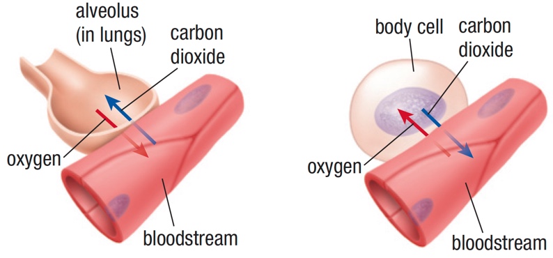
Back in the Structures Unit, you explored how the alveoli is the site of gas exchange between environment and our bodies, and then how blood carries carbon dioxide and oxygen to and from the cells.

In addition to gases, the blood carries other important components such as water, nutrients, and vitamins to our cells. Without the respiratory system, gas exchange would not occur. Without the digestive system, we wouldn’t be able to obtain the required nutrients. Without the circulatory system, these gases and nutrients couldn’t be transported to the cells where they are needed. Each system plays a role in the health of the organism, and the systems in our body are intimately interconnected, working to maintain homeostasis.(definition:The ability or tendency of a living organism, cell, or group to keep the conditions inside it the same despite any changes in the conditions around it, or this state of internal balance.) One system simply cannot function without the others. Although you will focus on the digestive, circulatory, and respiratory system in this course, it is important to understand that all of the eleven systems in our body play an integral role in the functioning of the others.

In Grade 10, you learned about the role of the different systems in the body. If you need a refresher, take a few minutes to look over this video that describes the main systems and how they are interconnected. Pay particular attention to the three systems we will explore in this activity to gain a better understanding of how they relate to one another.
The Fuel that Runs it All!
The digestive system is one of the systems this activity will cover in detail. Without nutrients, the digestive system wouldn’t function. Without the energy generated using the food you eat, none of the other systems would function either. Unfortunately, we do not have the ability to manufacture our own food through photosynthesis like our autotrophic friends. Instead we need to consume our food to get the raw materials our body needs.
You will look at the main macronutrients(definition:A substance required in relatively large quantities for growth, energy, and health.) in the Action section and see how each is processed through the digestive system. First, let’s take a few minutes to think about how we obtain these nutrients.
Canada’s Food Guide has an interactive website that allows you to create your own personal food guide. Take a few minutes to go through the process by clicking on the image below. You will see how these nutrients are used by your body in this activity.
Ready to Go!
The food guide should help you see the importance of macronutrients to the health and fueling of your body. The digestive, circulatory, and respiratory systems are amazing systems on their own, and even more amazing when you see them working in concert with each other. This activity will look at each of the systems, and then we will investigate how human interactions and choices have an impact on them.
You are now ready to look at each system in detail.

ACTION
Digestive System
Some people say that food is just feces(definition:Waste matter discharged from the colon after food has been digested.) in waiting, but it is what the food does as it goes through your system that is important. Before we look deeper into the anatomy and physiology(definition:The branch of biology that deals with the normal functions of living organisms and their parts.) of the digestive system, it is important to look at the main macronutrients that we need to survive. After all, our need for these nutrients is why we have a digestive system in the first place!
The Six Essential Nutrients
Carbohydrates, proteins, lipids, water, vitamins, and minerals are the building blocks of life. Without these essential nutrients, we would not survive. We need to consume greater amounts of the first three - carbohydrates, proteins, and lipids - so they are called macronutrients.(definition:Nutrients needed in large quantities.) Vitamins and minerals, on the other hand, are called micronutrients.(definition:Nutrients needed in small quantities.) Water is in a category by itself. You will learn more about the role of water in Grade 12 Biology.
Macronutrients come as both larger molecules, called polymers,(definition:Molecules made of many smaller molecules of the same kind called monomers.) and smaller ones, called monomers.(definition:A small molecule that reacts with a similar molecule to form a larger molecule.) Polymers are made of many monomer molecules joined together. Because different macronutrients are digested in different parts of the digestive system it is important to get to know the names of these monomers and polymers.

As you go through the information about the essential nutrients, it is important to concentrate on the monomers that make up each macronutrient listed. You will then be able to follow carbohydrates, proteins, and lipids from polymer to monomer as they go through the process of digestion. In addition, be sure to determine if the macronutrients are used for energy or growth. Some macronutrients are used for both. This information can then be included in the digestion organizer you will create in an upcoming task.
Carbohydrates
Carbs are the molecules that some people love to hate. Carbohydrate(definition:Biological molecules made of carbon, hydrogen, and oxygen usually in a ratio of 1:2:1.) reduced diets are all around us, but carbs are the main source of energy for your body. So, although there may be a benefit of reducing certain forms of carbohydrates, they cannot be eliminated.
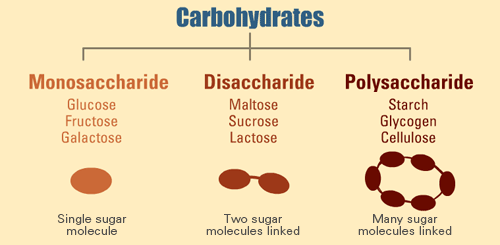
There are a number of different forms of carbohydrates: monosaccharides,(definition:The most basic unit of carbohydrates.) disaccharides,(definition:A sugar composed of two monosaccharides linked together.) and polysaccharides.(definition:Any carbohydrate formed by repeating units linked together.)
Monosaccharides, or simple sugars, such as glucose, are easy to digest. They are a source of energy for most living things. Disaccharides such as sucrose, better known as table sugar, are made of two monomers linked together. Polysaccharides contain many monomers linked together to form large molecules. These include starch, the carbohydrate storage molecule in plants, and glycogen, the carbohydrate energy storage molecule in animals.
Extension: Carbohydrate Structure
This video goes more deeply into the structure of the different types of carbohydrates and their various uses beyond energy sources or storage molecules.
Proteins
Proteins(definition:A molecule composed of amino acid monomers linked together by peptide bonds.) are large, complex molecules that play many critical roles in your body. They are involved in chemical reactions, transport, and defence. Therefore, they are an important structural molecule. Some proteins act as hormones,(definition:A chemical signal or messenger molecule that circulates through the body to coordinate cellular functions.) which are chemical messengers within your body.
Proteins are polymers made up of amino acids(definition:Monomers of proteins, each with a side chain structure unique to that amino acid.) linked together by peptide bonds.(definition:A chemical bond formed between amino acids.) Each amino acid has its own unique properties, and it is the combination of the amino acids and their properties that determines the function of the protein. There are 20 different amino acids. Most can be made by the body, but nine are essential and therefore need to be consumed.
Proteins can be used by the body as an energy source, but this usually only occurs if there aren’t any carbohydrates readily available. All the enzymes that control the various reactions within your body are proteins. In addition, proteins provide structure and support for cells and tissues.
Lipids
Lipids(definition:A group of biological molecules that include fats, oils and some steroids.) also play many important roles in your body. Not only are they a source of concentrated energy and a major form of energy storage, they are also the main component of cell membranes. In addition, some lipids act as a special form of hormones called steroids(definition:A lipid-based hormone.).
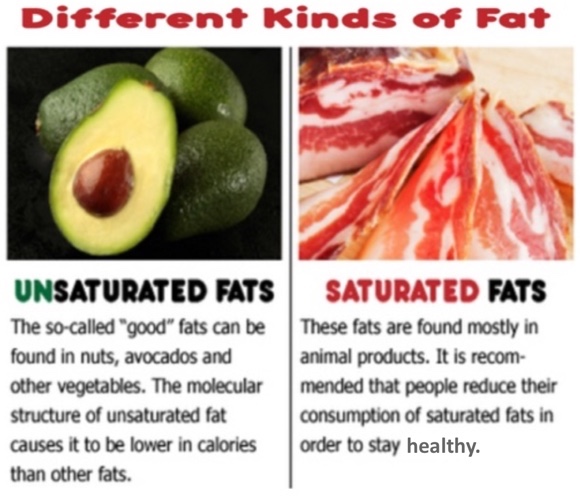
Fats and oils are lipids, and often get a bad rap. Although moderation is the key to all things we consume, lipids are an important part of our diet. Fats and oils are large molecules called triglycerides(definition:A lipid composed of three fatty acid chains and one glycerol molecule.). They are composed of three fatty acids and one glycerol molecule. Some fatty acids, such as the Omega-3 fatty acids, cannot be produced in the body and therefore must be consumed. This is why they are called ‘essential’ fatty acids.
You have most likely heard the terms saturated(definition:A fat which there are no double bonds between carbon atoms.) and unsaturated(definition:A fat which there are double or triple bonds between carbon atoms.) fats. Saturation simply means that no additional hydrogens can be added to the molecule. The following image compares the two forms of fat.
Extension: Biological Molecules
The following video will give you some additional detail on carbohydrates, proteins, and lipids. It emphasizes the importance of each in an entertaining way.
Water, vitamins, and minerals are also needed by the body. These three components will be discussed below:
Water
Your body is 60% water on average. Many chemical reactions that occur within your body require water. It is used to maintain your blood volume and also plays an important role in keeping your body cool in hotter weather.
Vitamins

Vitamins(definition:Organic compounds essential for normal growth and nutrition and are required in small amounts.) are not needed in large amounts in the body, but they are crucial for growth and the regulation of many cell functions.
As with macronutrients, some vitamins are labelled as ‘essential’, since the body cannot produce them on its own. Examples include Vitamins B and C. Although most of the vitamins we need are obtained through the food we eat, some can be produced by our body. A prime example is Vitamin D, which is formed when you are exposed to sunlight. Vitamin K is produced by bacteria that live in your large intestine, and Vitamin A is produced from beta-carotene. Beta-carotene is a chemical found in foods such as carrots and liver.
Minerals
Minerals(definition:A chemical element required as an essential nutrient by organisms to perform functions necessary for life.) are ions of elements that are found on the periodic table. Most are only needed in trace amounts, such as zinc and copper, but other minerals play a larger role in the functioning of our systems. Sodium is involved in muscle contraction and nerve pulses, while calcium and phosphorus play an important role in bone formation.
One of the most important minerals in our body is iron. Iron is part of the compound hemoglobin,(definition:A protein of red blood cells that contains iron and carries oxygen from the lungs to the tissues and carbon dioxide from the tissues to the lungs that is found in red blood cells.) which is found in red blood cells. The O2 from the gas exchange process binds to the iron component of hemoglobin and is then carried through the blood to the cells.
Let’s Get Down to Business!
Now that you know what the body is trying to obtain through digestion, it is time to look at the actual process. This video will serve as an overview of the anatomy and physiology of the digestive system. Each aspect will be explored in detail later in this activity. While you watch, keep an eye out for the various macronutrients and how they are broken down into their subunits, or monomers, in the process.
You have most likely heard the term, metabolism.(definition:The set of chemical reactions that occur within a living organism that are needed to sustain life.) This is a general term that covers all the chemical reactions that occur in your body to keep you alive. In the digestive process, the chemical reactions break down larger molecules into smaller subunits. This act of breaking down food is called catabolism.(definition:The metabolic reactions that break down large molecules into their subunits.) For example, proteins will be catabolized into amino acids.
Your body then takes the products of catabolism and, using energy, puts the subunits obtained together into larger molecules that the body needs. These reactions are called anabolism.(definition:The metabolic reactions that use energy to produce larger molecules from smaller subunits.)
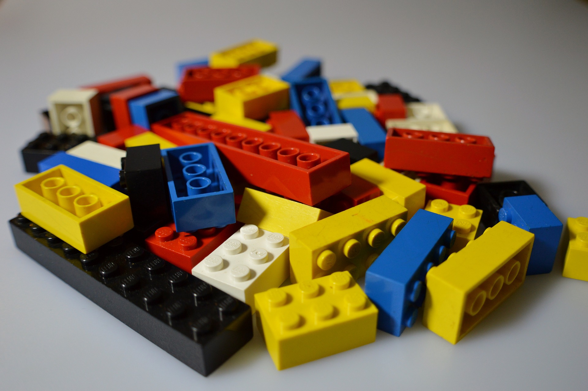
Think of metabolism as a child playing with Lego bricks. First, she will take apart any earlier made structures in order to separate the individual bricks (catabolism). Using these individual bricks, she will then create new structures (anabolism). See how much fun digestion can be? Until you step on a Lego, that is.
 Digestive System Organizer
As you go through the process of digestion, you will come across a number of structures and their associated functions.
It would be a good idea to colour code information about the three macronutrients. In this way, you will be able to trace the fate of each nutrient from polymer to monomer! Circling the vitamins, minerals, and water when they appear will help you keep visible track of all the nutrients throughout the digestive process.
How the Digestive System Provides the Body with Nutrients for Energy and Growth| Structure | Function | Chemicals Involved |
|---|---|---|
|
Mouth Includes teeth, tongue, and salivary glands. |
|
|
| ... | ... | ... |
Four Stages of Digestion
Digestion as a whole is actually a series of events:
- Ingestion - the taking in of nutrients;
- Digestion - the breakdown of food into smaller subunits;
- Absorption - the transfer of the digested nutrients into the bloodstream; and
- Egestion - the removal of waste products of digestion from the body.
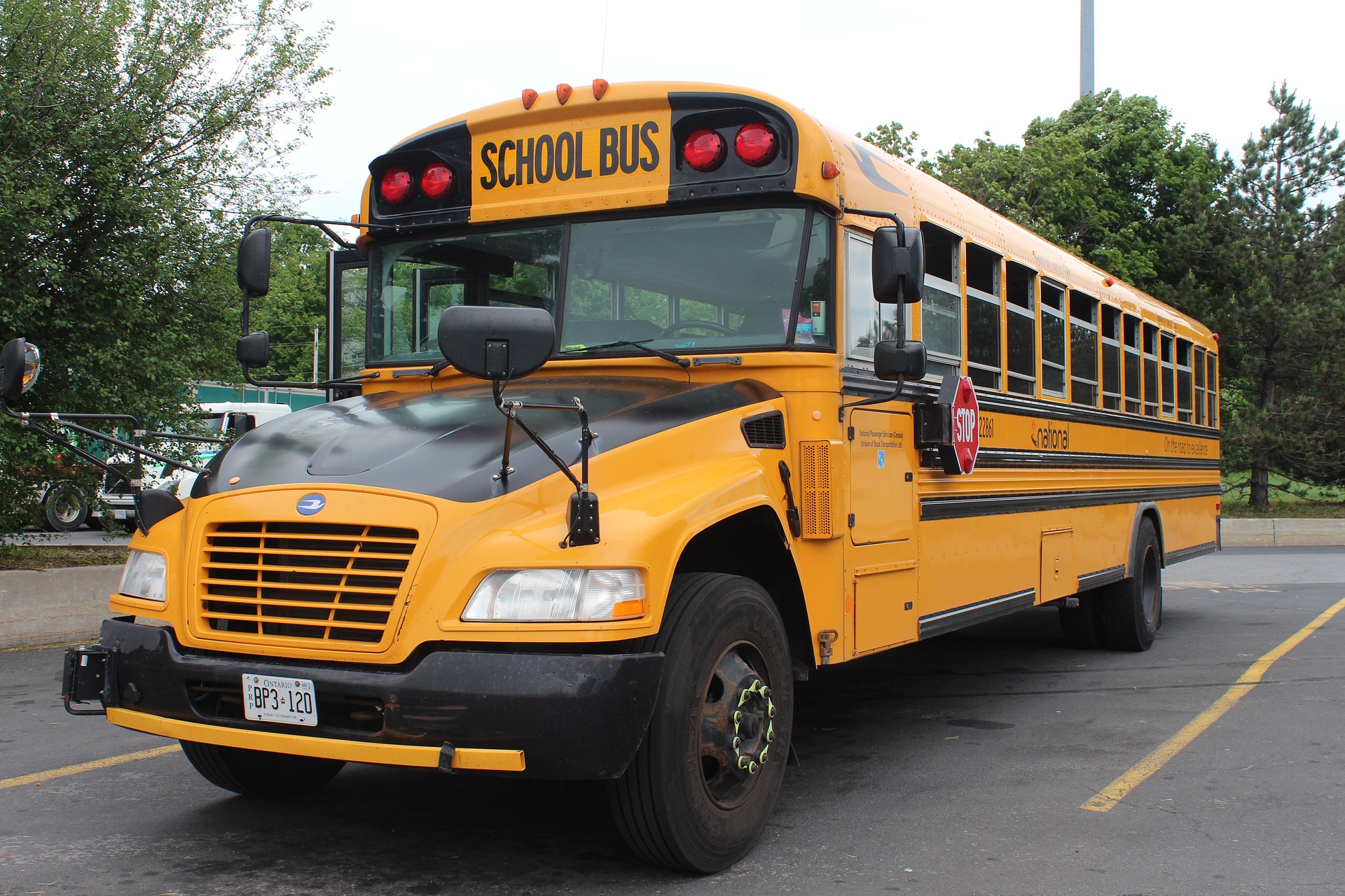
All these events occur within the gastrointestinal tract,(definition:A tube that extends from the mouth to the anus in which digestion occurs.) or ‘GI’ tract. In an adult human, this GI tract can be 7 - 9 metres long. That is almost as long as a full size school bus!
As you know, digestion is the physical and chemical breakdown of food into smaller molecules. The physical aspect of digestion occurs through the mechanical action of your body such as chewing with your teeth and mixing and moving the food with your muscles. Chemical digestion uses chemicals such as enzymes(definition:Proteins that speed up chemical reactions.) to break down the food.
Stage 1: Ingestion
Ingestion basically covers the act of putting food in your mouth and then swallowing it. This ingestion of food may seem uneventful, but lots is going on in this stage. The process of digestion occurs mainly in the next stage, but digestion actually starts as soon as food enters your mouth. Your teeth break up the food into smaller pieces, which is physical or mechanical digestion.(definition:The physical process of preparing the food for chemical digestion. It involves chewing, mixing, and churning.) Even thinking about eating stimulates the salivary glands to secrete saliva, which serves a number of purposes. Mucus in the saliva acts like a lubricant to help the food slide down your throat. The saliva also contains the enzyme amylase,(definition:A digestive enzyme produced largely by the pancreas and salivary glands that converts starches to sugars.) which breaks down starch into its simple sugars, like monosaccharides. This is the true start of chemical digestion.(definition:Where food is broken down by the action of chemical agents and enzymes.) In addition, the food mixes with the saliva and allows the taste buds of your tongue to do their job.
Try This!
Put an unsalted cracker into your mouth, and start chewing. Don’t swallow right away, but instead pay attention to the taste. At some point, you will start to detect a sweet taste. Starch in the cracker is not sweet, but once the enzyme amylase starts to break it down into simple sugars, you can taste the sweetness!
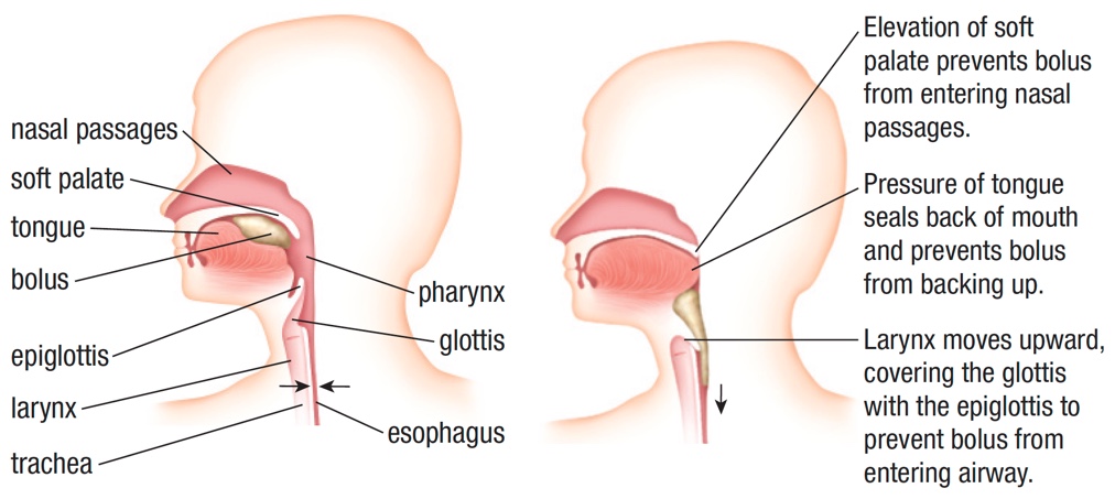
Once the food is chewed and mixed with saliva, this mixture is now known as a bolus(definition:Food that has been chewed and mixed with saliva.). The tongue will now push the bolus to the back of the throat in order to swallow it.
When you swallow, a small flap of tissue called the epiglottis(definition:A thin, triangular plate of cartilage at the base of the tongue that covers the glottis during swallowing to keep food from entering the trachea.) closes the trachea so that the food does not go into your lungs. Have you ever started to choke when you start to laugh while eating? This is because the act of laughing prevents the epiglottis from sealing the trachea while you swallow. So, don’t laugh while you eat!
Once you swallow, the bolus makes its way to the stomach via the esophagus. Although gravity plays some part in this, it is the rhythmic, wave-like motion of the muscles that make up the esophagus that actually pushes the food down. This peristalsis(definition:Involuntary wavelike contractions of the muscles in the esophagus.) is why you can swallow food even if you are upside down!
Stage 2: Digestion
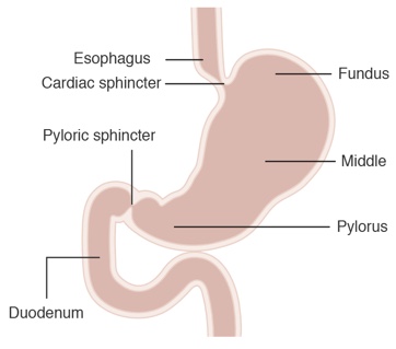
This is the stage where the real magic happens! The bolus is pushed down the esophagus and then into the stomach. The cardiac sphincter is a muscular ring at the top of the stomach that prevents material from going back up the esophagus. The pyloric sphincter is located at the bottom of the stomach, and controls the release of material into the next section of the system.
The stomach is composed of a number of layers. The innermost layer is called the mucosa,(definition:A lubricating membrane lining all body passages and cavities.) and it secretes gastric juices.(definition:A secretion of the gastric glands in the stomach that includes hydrochloric acid, pepsinogen and mucus.) The gastric juice contains the enzyme pepsinogen.(definition:A substance made by cells in the stomach. A precursor to pepsin.) This enzyme is active only in low, or acid pH environments, so that is where the acid comes in. The acid produced is hydrochloric acid, which is very strong. This acid converts the pepsinogen into its active form, pepsin.(definition:An enzyme that breaks down protein.) This enzyme breaks down the proteins in the food into their amino acid monomers. This acid is so strong, that it could actually burn through the stomach lining. The mucus in the gastric juice coats the stomach lining and protects it from the acid. This is one harsh environment! In fact, the amylase enzyme from the saliva that moves with the bolus is deactivated by the acidic conditions in the stomach.
Did You Know?
Other layers of the stomach are made up of muscles. These muscular layers contract and help mix the food with the gastric juices. Once mixed with the gastric juices, the bolus is now called chyme.(definition:A semi-liquid mixture of the bolus and gastric juices.)
This mixing by the muscular layers is considered mechanical digestion, while the action of the enzymes is chemical digestion.
There is More to the Story!
Although chemical digestion occurs within the stomach, a good amount of the chemical digestion also occurs in the duodenum,(definition:The first 25 to 30 cm of the small intestine.) which is the first section of the small intestine attached to the stomach. You will learn more about the small intestines soon.
Once the chyme is well mixed, the pyloric sphincter at the bottom of the stomach will relax and allow it to move into the duodenum a little at a time. Remember, the chyme will be very acidic, and this acid can damage the lining of the duodenum...this is where the pancreas comes into play.

The pancreas releases bicarbonate ions that are secreted into the duodenum. These ions neutralize the acid secreted by the mucosa of the stomach and the pH level goes up. Just as when the amylase moved into the stomach, the change in pH now deactivates the pepsin from the stomach gastric juice. Although pepsin is now out of the picture, proteins will continue to be digested by an another enzyme called trypsin(definition:One of the enzymes used to digest proteins.) that is also secreted by the pancreas. In addition, the pancreas will secrete additional amylase into the duodenum to replace the enzyme destroyed in the acidic environment of the stomach. This means that carbohydrate digestion will continue once again.
So far, you have seen how the carbohydrates and the proteins are broken down. What about the lipids? Well, the pancreas has another trick up its sleeve. Along with the ions and the other enzymes, the pancreas secretes the enzyme lipase(definition:Enzyme that breaks down dietary fats in human digestive system.) into the duodenum. The issue is that fats are in large insoluble globules, so the lipase cannot do its job properly. This is where the bile(definition:A greenish-yellow bodily fluid secreted by the liver and stored in the gallbladder, and discharge into the duodenum where it aids in the digestion of fats.) helps out. The liver produces bile, which is stored in the gallbladder until needed. This bile then enters the duodenum and emulsifies(definition:Breaks down fats into tiny droplets allow them to mix with other liquids.) the fat into tiny little droplets. These droplets increase the surface area of the fats, and allow the lipase to do its job of breaking down the fats into their monomers - glycerol and fatty acids.
Stage 3: Absorption
The small intestine has three main sections, the duodenum, jejunum,(definition:The section of small intestine between the duodenum and ileum. It is approximately 2.5m long.) and ileum.(definition:The final section of the small intestine. It is approximately 3.5m long.) Many minerals are absorbed by the duodenum. Macronutrients are absorbed in the jejunum, while some vitamins are absorbed in the ileum. Some water is absorbed by osmosis throughout the small intestine.

Now that the carbohydrates, proteins, and lipids have been broken down, it is time to absorb(definition:Take in a substance, such as a liquid or solute, across a cell membrane for example by diffusion or osmosis.) the nutrient monomers into the bloodstream so they can be delivered to where they are needed. The method of absorption depends on the molecules being absorbed. Some can cross membranes using simple passive transport such as diffusion that doesn’t require energy, while other, larger molecules may need energy and active transport to get them into the blood. No matter the process, the absorption will occur through the walls of the small intestine. The walls are highly folded to increase the total surface area of the intestine lining. The folds appear as little fingers called villi.(definition:Finger-like projections of the inner lining of the small intestine. Singular is villus.) To increase the surface area even more, the villi have even smaller projections on them called microvilli.(definition:Tiny projections found on the surface of villi. Singular is microvillus.) If you smoothed out all the villi and microvilli, the surface area of the small intestine would be about the size of a tennis court! All of this increased surface area allows the small intestine to absorb more nutrients in less time.
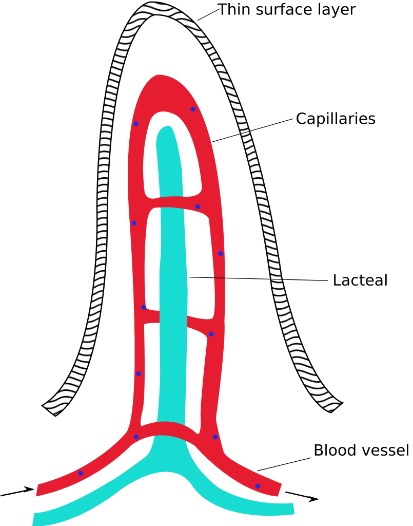
Each villus has a network of capillaries behind the thin outer layer. You will remember from Unit 1 that capillaries are small blood vessels in the circulatory system. All nutrient monomers, except for the breakdown products of lipids that are absorbed through the villi, will enter the bloodstream for transport around the body. The molecules from the digestion of fats will instead be collected in the lacteal(definition:A projection into each villus that collects the digested fats after absorption.) and then into the lymphatic system.(definition:A system similar to the circulatory system that transports a fluid called lymph around the body and to the bloodstream.) Later, it will be transported into the bloodstream. From the end of the small intestine, the material moves into the large intestine.
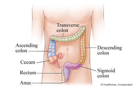
The large intestine is approximately 1.5m in length, but it has a greater diameter as compared to the small intestine. The large intestine connects to the small intestine a few centimetres before the end, creating a small pocket called the cecum. At the bottom of the cecum lies the appendix.
Did You Know?
The colon, or large intestine, has four sections: the ascending colon, transverse colon, descending colon, and the sigmoid colon. These sections are names based on their positioning and general structure.
Although most of the nutrients are absorbed in the small intestine, there is some absorption that takes place in the large intestine, as well. Most of the water is absorbed from the material that enters the colon through osmosis. More ions and vitamins are also absorbed into the body at this point. The remaining material is mostly indigestible wastes such as cellulose and some water to keep the material moving through the system. It is now called feces.
Stage 4: Egestion
The material is now close to the end of its voyage through the GI tract. At this point, all the nutrients and water have been absorbed from the food that was ingested. Once through the colon, these wastes are stored in the rectum, which is the last 20 cm of the GI tract. When enough wastes have collected in the rectum, the feces are eliminated through the anus.
The Incredible Journey!
You have just been introduced to a lot of new terminology and concepts. You were given an overview at the start of the digestion section, so a good way to end it is with a summary. In this video, the digestive systems of other animals is briefly discussed. The digestive system of animals is based upon the diet that they have, so there are some differences.
Now that you are a digestion expert, try your hand at this timed labelling interactive.
Digestion
That is a lot of information to digest! There are a good number of structures and chemicals needed to make digestion efficient. Evolution has made modifications along the way, and the digestive system found in animals, including humans, has changed as the environment and the available resources have changed.
Circulatory System
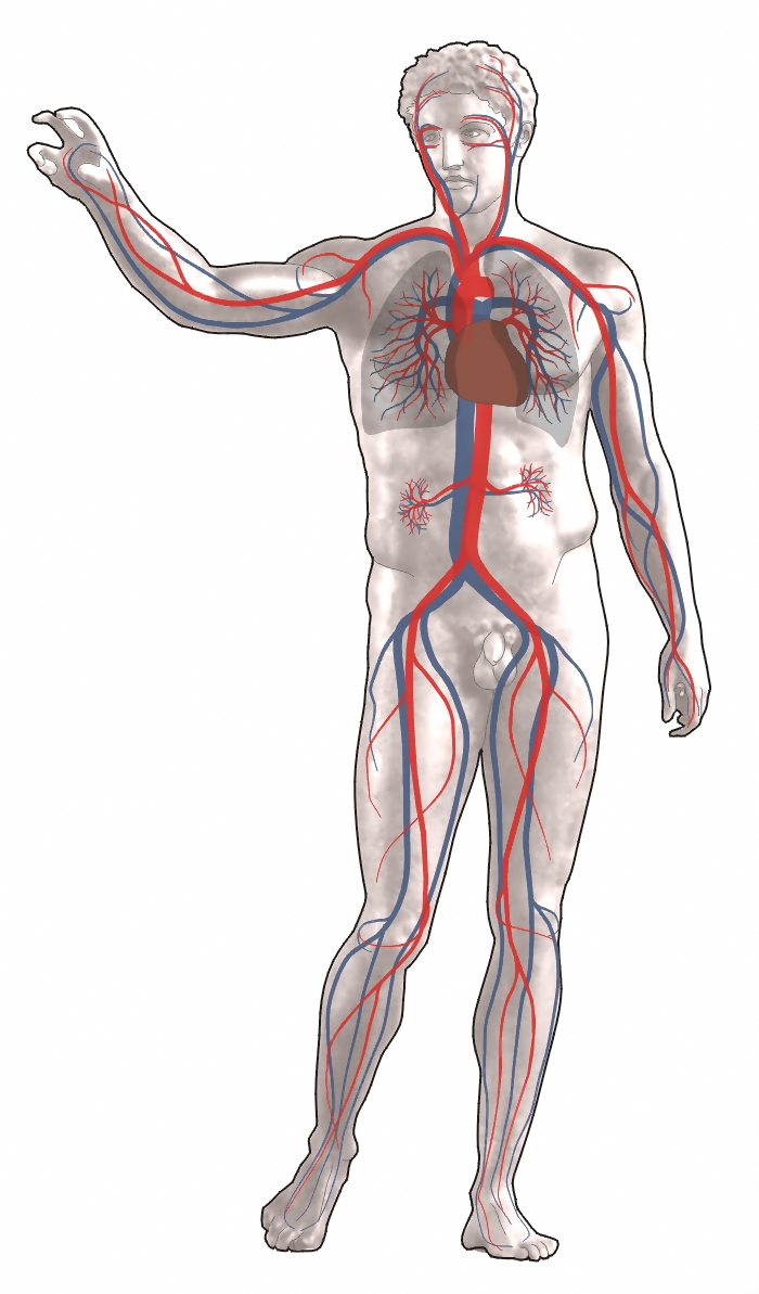
Now that the food has been processed through the digestive system, the next thing to do is to transport the resulting molecules to the rest of the body where they are needed. This duty falls to the circulatory system. This system is composed of the heart, blood vessels, and the blood, itself.
In Unit 1, you looked at the different forms of circulatory systems that various animals use. Less complex animals such as insects utilize an open circulatory system, where the blood bathes the tissues directly. More complex animals have closed circulatory systems, where the blood is contained in a series of vessels. Fish have a single closed system, where the blood travels in one loop throughout the body. Other more complex animals, such as humans and other mammals, use a double loop closed system. In this system, there are two loops, or circuits, and the blood travels through the heart twice as it travels through the body. It is this double closed circulatory system that you will look at in detail in this activity.

In humans and other mammals, you will find the pulmonary circuit and the systemic circuit. The systemic circuit(definition:The part of the circulatory system which carries oxygenated blood away from the heart, to the body, and returns deoxygenated blood back to the heart.) starts at the heart, which pumps oxygenated blood and nutrients to the tissues of the body. The blood then picks up the CO2 and other wastes from cell metabolism and this oxygen-poor blood returns to the heart.
The heart then pumps this deoxygenated blood through the pulmonary circuit.(definition:The part of the circulatory system which carries deoxygenated blood away from the heart, to the lungs, and returns oxygenated blood back to the heart.) The CO2 is then exchanged for O2 at the alveoli, and this oxygen-rich blood returns to the heart to start the entire cycle over again.
Circulatory System Organizer
Just as with the digestive system, you will come across a number of structures and their associated functions when exploring the circulatory system. Create a graphic organizer such as a chart with which to keep track of this information. The organizer can be of your own design, but here is an example:
How the Circulatory System Transports Substances that are Vital for Health| Structure | Function |
|---|---|
| Capillaries | Sites of oxygen and other nutrients transfer from the bloodstream to other tissues in the body and waste products from the cells to the blood. |
| ... | ... |
Your organizer should also include the structures of the heart.
Circulatory Overview
The following video explains the path that the blood takes through the system, and how the heart works. You will cover this information in detail in this section of the activity.
You Gotta Have Heart!
The heart truly is an amazing organ that beats an average of 70 times a minute. Just imagine doing anything 100,000 times a day! Often the heart is referred to as a basic pump, but it is actually a very complicated structure that requires precise timing and specialized structures for it to function efficiently and be able to pump approximately 5 L of blood per minute.
The heart is a muscular organ that is found just under the breastbone in the middle of your chest. When you look at diagrams of the heart, you will see that the left and right are backwards. When you look at a diagram, just imagine that you are looking at the heart of someone facing you. If you get this individual to raise her/his right arm, the raised arm would be to the left from your perspective.
Structure of the Heart

As the diagram illustrates, the walls of the right and left ventricle(definition:A chamber in the heart which forces blood into the arteries.) are very muscular, as well as the septum that separates them. The left ventricle has the thickest muscle, as it needs to pump blood to all parts of your body, right down to your toes, through the systemic circuit. The right ventricle doesn’t need to be as strong, as it pumps blood to the nearby lungs in the pulmonary circuit. The walls of the right and left atria(definition:Each of the two upper cavities of the heart from which blood is passed to the ventricles. Singular is atrium.) do not need to be as muscular, as they just have to pump the blood they receive from the veins into the ventricles.
You can see that there are valves located between the atria and the ventricles, and again between the ventricles and the arteries. These valves ensure that the blood flows in only one direction when each chamber contracts. The diagram shows two names for each valve, and both terms are commonly used.
Did You Know?
This echocardiogram(definition:A visual display of the heart produced by ultrasound waves.) shows the heart valves in action:
Directing Traffic
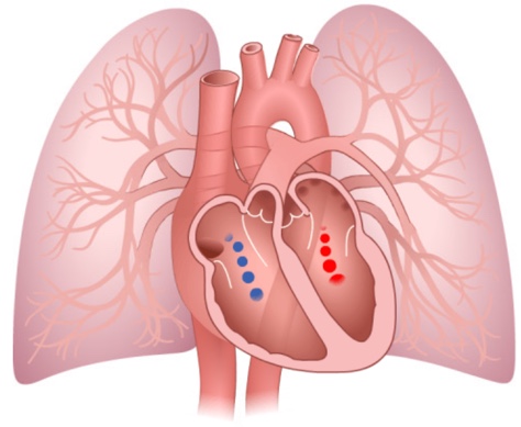
It is the heart that directs the blood to flow through the proper circuit. The deoxygenated blood needs to be routed to the lungs through the pulmonary circuit, while the oxygenated blood needs to travel through the systemic system to the body tissues. Without the different chambers and valves of the heart, the system wouldn’t be efficient.
Click on the screenshot below to see how the four chambers of the human heart work in concert to pump the blood to where it is needed. Select the Contraction tab at the top and then use the arrow buttons to move through the animation.
Here is the path that the blood follows through the body, starting from the body tissues. Don’t forget to include the structures of the heart on your system organizer! Click on the image below for an animation of the blood flow through the heart. Select the Blood Flow tab at the top and then use the arrow buttons to move through the animation.

The following video shows you another view of the movement of blood through the heart. Pay attention to how the valves between the chambers and the major vessels function as the heart beats.
The step by step route of blood flow through the heart is detailed below. Refer to the labelled image of the heart to follow the flow:
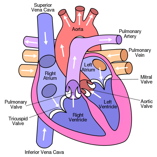
- Deoxygenated blood from the body enters the right atrium from the superior and inferior vena cava.
- From right atrium, blood goes through the tricuspid valve to the right ventricle.
- From the right ventricle, blood goes through the pulmonary semilunar valve to the right and left pulmonary arteries.
- From the pulmonary arteries, the blood flows to the lungs.
- From the lungs, the now oxygenated blood is returned to the heart through the left and right pulmonary veins.
- From the pulmonary veins, blood flows into the left atrium.
- From the left atrium, blood flows through the mitral valve into the left ventricle.
- From the left ventricle, it goes through the aortic valve into the aorta.
- From the aorta, the blood flows through the arteries to the body tissues.
- The deoxygenated blood then flows through the veins and back into the superior and inferior vena cava and starts the cycle again (go back to Step 1).
Do you think you can trace the flow of the blood through the heart? Using good self regulation(definition:I am able to set my own goals in achieving mastery of the blood flow through the heart.) skills, try this interactive to find out!
Flow
Rhythm Control
It is obvious that the timing of the ventricle and atria contractions are vital to proper blood flow through the system. How are these contractions controlled? Refer to the animation at the start of the Directing Traffic section for a reminder of the heart contraction process.
The interesting bit is that the heart does not need external stimulation to beat. Instead, it has special myogenic muscles(definition:Muscle fibres that can contract without stimulation from a nerve cell.) that can contract without being stimulated to do so by an external nerve cell.
The heart essentially has its own internal nervous system that is controlled by a cluster of cells called the sinoatrial node,(definition:A group of cells located in the wall of the right atrium of the heart that generates electrical signals.) or SA node. This SA node acts like a pacemaker and controls the rhythm of the heart. The SA node, located in the right atrium, generates an electrical signal that travels through the walls of the atria making them contract. The signals then continue on and stimulate another cluster of cells and fibres found in the wall between the right atrium and the right ventricle called the atrioventricular node,(definition:A small mass of fibres located in the wall of the right atrium of the heart that receives electrical impulses from the SA node and directs them to the walls of the ventricles.) or AV node. This AV nodes acts like a signal repeater and sends the impulse that originated from the SA node down the Purkinje fibres within the ventricle walls and making them contract.
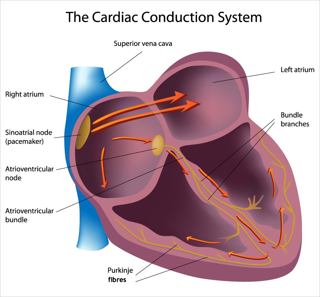
An electrocardiograph,(definition:The image produced by a test that records the electrical activity of your heart.) or ECG, detects the electrical pulses produced by the heart so that medical professionals can have an image of the rhythm to ensure the heart is beating properly. This ECG shows three cycles of a heart:
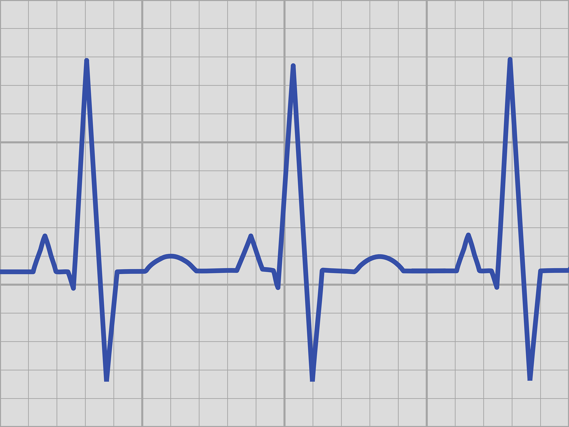
Click on the image below for an animation explaining the peaks that appear on the ECG. Select the ECG tab at the top and then use the arrow buttons to move through the animation.
Even though the heart has its own system to control its rhythm, this isn’t to say that external factors cannot influence the heart rate. Receptors in your medulla oblongata(definition:Located in the brainstem and responsible for involuntary functions such as breathing and heart rate.) detect changes in blood gases and impact your heart and respiratory rate. You will learn more about this in the next activity. If your body experiences stress of some sort, the nervous system can signal an increase in the heart rate in order to supply the muscles with oxygen at an increased rate. This is important if you need to fight off the danger or run from it! This ‘fight or flight’ reaction is very important for the survival of our species!
Blood Vessels

Obviously, the pump is a very important part of the system, but the blood wouldn’t get to where it was needed without the series of blood vessels throughout your body.
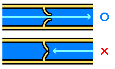
In Unit 1, you saw the specialized structures involved with the veins and arteries. Here is a side-by-side comparison of the two vessels to jog your memory:
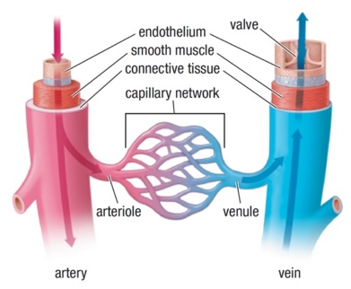
As you can see in the comparison of the two types of vessels, many of the larger veins have valves where the arteries do not. The strong left ventricle produces enough pressure through the arteries for the blood to get to the tissues and cells. The pressure in the veins is considerably lower, and that blood has to get from the feet all the way back up to the heart against gravity. Not an easy task!
This is where the valves and the skeletal muscles come into play. The muscles will contract and squeeze the blood up through the veins, and the valves will prevent the blood from flowing back down when the muscles relax.
Try This!
Blood Composition
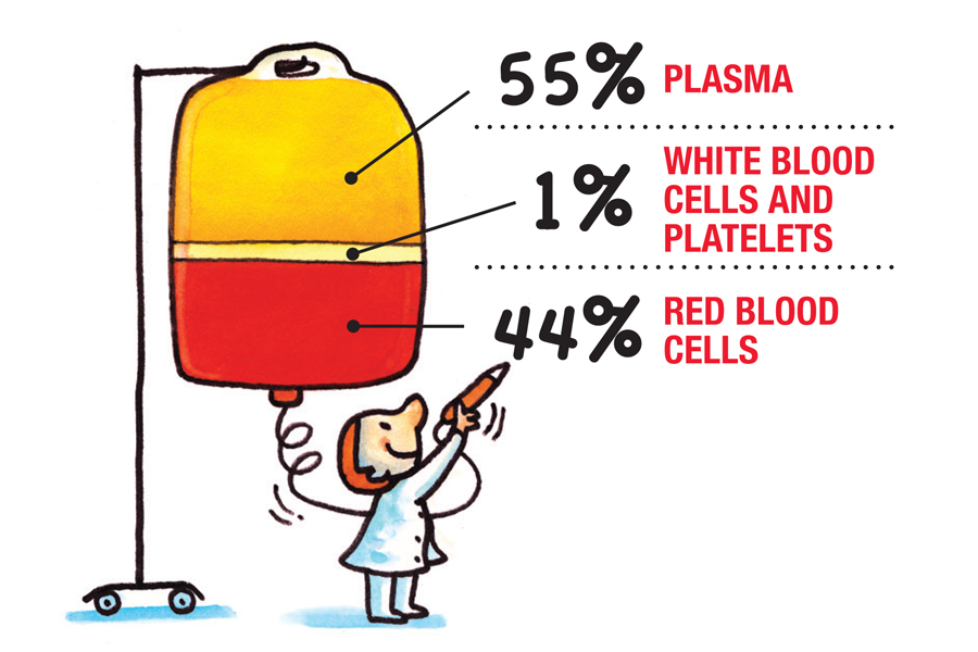
As you know from earlier activities, blood is considered a connective tissue and connects all the organ systems of the body by transporting oxygen, nutrients, hormones, etc., and removing wastes from these organs. Blood has four main components: plasma, red blood cells, white blood cells, and platelets.
Adding to Your Circulatory Organizer
Using your research skills, determine the basic structure and functions of each blood component and add it to the circulatory organizer you started earlier.
As you explore, think about how each component could impact other systems of the body.
This short video provides a good summary of blood composition and the roles that each component plays.
So far you have looked at the fuel coming into your body, the digestion of this fuel into useful components, and then the transport of these components to the tissues and cells that need them. The wastes produced by the metabolic reactions within your cells now need to be removed from the body. In Grade 12, you’ll learn more about the role of the kidneys and the urinary system in removing metabolic wastes. For our purpose here we’ll focus on CO2 waste. Cue the respiratory system!
Respiratory System
In the Structures Unit, you explored gas exchange in animals. The respiratory system used depends on the environment in which that animal lives. You saw that fish are able to capture O2 from the water through gills, while insects have special tubes that allow gases in and out of the body.
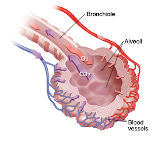
When you looked at the gas exchange in more complex animals such as humans, you saw that the gases diffused from the environment into the body, and vice versa, in internal structures called alveoli.(definition:Any of the many tiny air sacs of the lungs which allow for rapid gaseous exchange.)
So, you know how the gases are exchanged, but how does the air get to these alveoli?
This short video is a nice overview of respiration. Take a few minutes to see the process in its entirety. Then you will look at the structures involved in more detail.
Respiratory System Organizer
Just like with the circulatory system, you will come across a number of structures and their associated functions when exploring the respiratory system. Create a graphic organizer such as a chart on which to keep track of this information. The chart can be of your own design, but here is an example:
How the Respiratory System Performs Ventilation and Gas Exchange| Structure | Function |
|---|---|
| Nose (nasal cavity) | Warms, moisturizes, and filters air entering the body before it reaches the lungs. |
| Mouth (oral cavity | Secondary external opening for the respiratory tract. |
| ... | ... |
As mentioned, gas exchange is just one aspect of respiration. The air first needs to get into the lungs and to the alveoli for the exchange to occur. This process is known as ventilation.(definition:The exchange of air between the atmosphere and the lungs achieved by the physical act of breathing.)
Ventilation
In order to breathe, a pressure differential must occur. Air moves from an area of high pressure to low pressure. In order to inhale, the pressure inside the lungs must be less than that of the outside atmosphere. To exhale, the pressure in the lungs must be greater than that of the atmosphere. Your body creates this pressure differential by mechanical means.
This mechanical ventilation is achieved through the contraction and relaxation of specialized muscles associated with the respiratory system. These are the diaphragm(definition:The muscle that separates the chest cavity from the abdomen and is the main muscle involved with breathing.) located beneath the lungs, and the intercostal muscles(definition:Muscles located between the ribs that contract and relax to aid breathing.) located between the ribs.
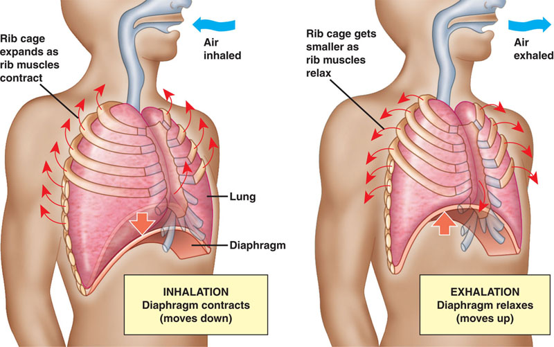

Inhalation is achieved by increasing the volume of the lungs. The greater the volume, the lower the pressure. When you breathe in, the diaphragm contracts and pulls down. At the same time, the intercostal muscles contract and push the ribs upward and outward. Place your hands on your chest and take a deep breath. You will feel your thoracis cavity(definition:The chest cavity.) expand as the muscles contract.
Now that the pressure is reduced within the lungs, the air will be pushed in by the higher pressure of the surrounding atmosphere. Through this process, the air can make its way into the alveoli for the gas exchange.

Exhalation is the reverse of this process. The intercostal muscles and the diaphragm relax, causing the volume of the lungs to reduce. This reduction in volume creates a higher pressure, and the air is pushed out of the lungs to the lower pressure area of the surrounding atmosphere.
This real MRI(definition:Magnetic Resonance Imaging. A diagnostic technique that uses magnetic fields and radio waves to produce a detailed image of the body's soft tissue and bones.) shows the volume of the thoracic cavity changing due to the action of the intercostal muscles and the diaphragm. The diaphragm is just above the liver and the beating heart is visible in the centre of the image.

There are thin, moist membranes called pleural membranes(definition:Two layer membrane that lines the chest cavity and the outside of the lungs to reduce friction between them.) that line the chest cavity and the outside of the lungs. These membranes reduce friction between the chest wall and the lungs while breathing.
Respiratory Structures

When you inhale, the air will rush through your nose and mouth and down the trachea(definition:The tube leading from the mouth toward the lungs.) to the lungs. Each component plays an important role in the respiration system.
Although you can breath through your mouth, the nose is the main entrance point for air under normal circumstances. When air passes through the nostrils, fine hairs and sticky mucus will help to capture small particles such as dust and bacteria. The air is then moistened and warmed in the nasal cavity to prepare it for its journey.
The air then passes through the pharynx(definition:Cavity behind the nose and mouth, connecting them to the esophagus.) and into the trachea. If you recall the digestive system, you will remember that the epiglottis covers the trachea when you swallow food to prevent solid materials from entering the lungs. When breathing, the epiglottis remains open to allow the air to pass through. At the top of the trachea is the larynx,(definition:The upper part of the trachea in humans, in which the vocal cords are located.) which houses your vocal cords.
The trachea contains multiple c-shaped bands of cartilage(definition:Flexible connective tissue.) along its length to prevent the trachea from collapsing during the constant change in pressure. In addition, the trachea is lined with tiny hairs called cilia and cells that produce mucus. The mucus will capture much of the small particulate matter that gets passed along the nasal passages and is then removed through the sweeping action of the cilia.
From there, the air branches to the left and right lung through the bronchi.(definition:Major air passages of the lungs that diverge from the windpipe.) These bronchi then branch into smaller bronchioles(definition:Any of the minute branches into which a bronchus divides.) and terminate in the tiny alveoli where the gas exchange occurs.
When you exhale, the air carrying the waste CO2 from cellular respiration is pushed out to the atmosphere and the cycle repeats itself...about 17,000 to 30,000 times a day!
Now that you have made it through the digestive, circulatory, and respiratory systems, it is an excellent time to use your good organization(definition:I am able to review the system charts and ensure all details are included.) skills to go back to fill in any missing details you may have omitted on your system organizers.
Hopefully, you now have a good understanding of the three systems explored in this activity. Continue to practice good self regulation(definition:I am able to monitor my own goals in achieving mastery of understanding how human systems function.) skills as you use your organizers to respond with great details to the following questions. If you have trouble with any of them, be sure to go back and review the material within the activity.
 Systems Questions
- Why is it important that the stomach secretes pepsin in its pre-enzyme or unactivated form, called pepsinogen?
AnswerPepsin is an enzyme that digests proteins. Since protein is a major component of our tissues, there would be a great risk of self digestion if the enzyme was active prior to being released into the stomach. Once secreted, the layer of mucus protects the stomach lining from self digestion, so the pre-enzyme can then be activated.
- Why is our digestive system is so complex? In other words, why can’t one organ do all the functions of the digestive system?
AnswerThe nutrients that we eat need to be broken down into their monomers. Each major nutrient, carbohydrates, proteins, and fats, requires specific chemicals and enzymes to break it down. Mechanical digestion is also needed to ensure proper mixing of these chemicals and the food. The system also needs a large surface area in order to absorb all the nutrients into the bloodstream. A simple, straight tube wouldn’t retain the food long enough in order to provide efficient absorption.
- Compare and contrast ventilation with gas exchange.
AnswerVentilation is the process of bringing air into the body from the atmosphere. The contraction and relaxation of the diaphragm and intercostal muscles changes the volume of the chest cavity in order to provide pressure differentials. Once the air is in the alveoli, oxygen can then diffuse into the bloodstream. The waste product, carbon dioxide, moves from the blood to the interior of the alveoli. Once this gas exchange occurs, the ventilation process pushes the waste gas out of the lungs.
- Explain why your respiration rate increases during exercise.
AnswerAs you exercise your muscles require additional energy. Energy is produced through cellular respiration. The higher demand for energy due to the increased muscle contractions means that a higher level of carbon dioxide is produced. Some of the carbon dioxide is converted to carbonic acid in the blood, which lowers the pH. Receptors detect this change, and the medulla oblongata signals the respiration and heart rate to increase. The increased respiration clears the blood of the carbon dioxide quicker, and also supplies the increased demand for oxygen by the muscle cells.
- Compare and contrast the pulmonary and the systemic circuit, and indicate the advantage of having a four chambered heart.
AnswerThe pulmonary circuit is the loop that pumps deoxygenated blood from the heart to the lungs in order for gas exchange to occur. The oxygenated blood then returns to the heart. The systemic circuit then delivers the oxygenated blood to the different parts of the body, and then returns the deoxygenated blood to the heart so it can go through the pulmonary circuit again. Having a four chambered heart prevents oxygenated blood from mixing with deoxygenated blood. This makes the delivery system of oxygen more efficient.
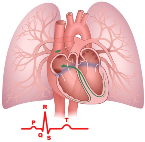
- Describe how the cardiac conduction system controls the rhythm of the heart.
AnswerThe cluster of cells known as the SA node or ‘pacemaker’ is located in the upper wall of the right atrium. The SA node generates an electrical signal that travels through the walls of the atria making them contract. This contraction pushes blood from the atria into the ventricles. The signal from the SA node is then detected by the AV node located in the bottom wall of the right atrium. Once detected, the AV node acts like a signal repeater and sends the electrical impulse along the Purkinje fibres that run through the ventricular walls, making them contract and push the blood out of the heart through the arteries.
What Does it All Mean?
Hopefully you noticed that one system cannot function properly without the others. All the systems in the body are intimately linked with overlapping structures that interact together.
As mentioned in Unit 1, the processes that occur in the body rely on specialized structures, and each has a unique role to play. In each case, form equals function, with structures evolving into a form that best fits the current conditions in which we live. All the systems in the body work in concert. If there is an issue in one, the others will also feel the effect. The next activity will look into this interconnectedness in greater detail.
CONSOLIDATION
Taking it All Apart!
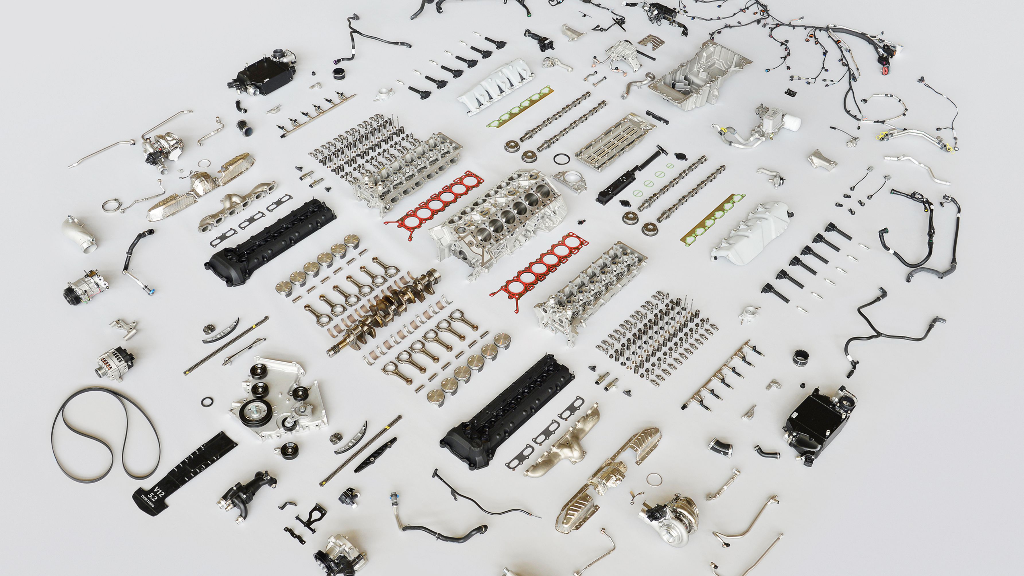
Throughout this activity, you have looked at the systems in the body and how they interconnect. By putting it all together, you have gained a better understanding of how one system relies on the others. Well, now it is time to take them apart!
Putting it All Together Again!
Millions of years of evolution has resulted in the the incredible machine you see whenever you look in the mirror. Each system does its part to ensure its efficiency.
The systems explored in this activity are amazingly complex, yet work very efficiently. Unfortunately, various factors such as inheritance of traits, disease, and environmental conditions (which are often influenced by human interactions) can mean that these systems will not work optimally. The impact on these systems by various diseases and disorders, along with the diagnostic tools used to monitor these systems, will be investigated in the next activity.