Biological Molecules
Putting it Together: Cellular Structures
MINDS ON
Some Key Terms
Look for these terms in this Activity. You should practice using them as you compose your responses to the upcoming tasks. In particular, make sure to incorporate these terms in the Consolidation task.
Realize, though, that this list is not exhaustive so you should look for opportunities to include other relevant terminology related to cellular structures.
|
micrometer |
electron microscope |
X-ray crystallography |
|
mitochondria |
chloroplasts |
nucleus |
|
cell membrane |
lysosome |
vacuole |
|
ribosome |
endoplasmic reticulum |
Golgi apparatus |
|
cytoskeleton |
control group |
|
Size in Cells
In our daily lives we develop an understanding of the sizes of different objects we see and use. It’s easy for you to compare two objects, say a pillow and a smartphone. With a bit of thought, you could confidently estimate how many times bigger the pillow is compared to the phone or even your room.
It’s the same thing with cells. Different components of the cell have different sizes compared to one another. It is possible to estimate how many mitochondria could fit in the diameter of a nucleus. In human cells, it’s about 6. Understanding this cellular scale will be important as we start to look at different technologies for viewing parts of the cell.
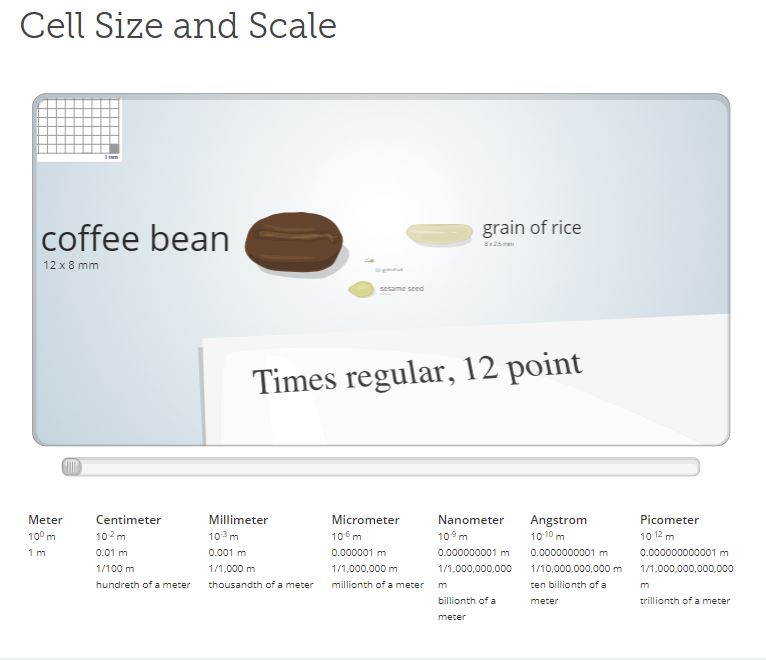
Start by exploring this interactive from Genetic Science Learning Centre showing the sizes of different objects, from a 12 mm long coffee bean to a 340 pm carbon atom. From our perspective, both these objects are small. Click on the graphic below, then drag the grey marker across the slider to start exploring.
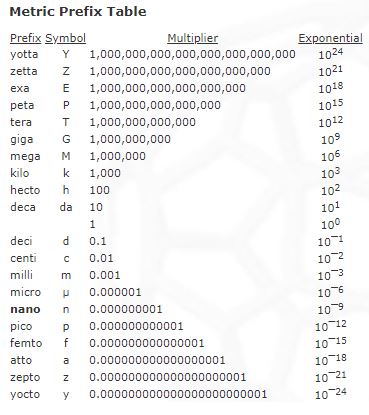
You might find the following table helpful.
 Example
Let’s take a moment to see how much bigger a coffee bean is compared to a carbon atom. In the interactive we see that a coffee bean is 12 mm in size. In other words, it’s 12 x 0.001 m. The carbon atom, on the other hand, is 340 pm which is 340 x 0.000000000001 m. So to compare how much bigger a coffee bean is compared to a carbon atom we divide the larger object by the smaller object:

 Relative Sizes of Cellular Components
Using the interactive, make a chart or scale to show how many times each of the important structures shown below is larger than a water molecule. Add this to your Portfolio as you will use this information later.
Important biological structures include:
- monomers of macronutrients (like glucose or amino acids)
- structural biological molecules (like phospholipids)
- larger biomolecules (like hemoglobin and ribosomes)
- viruses, like HIV
- vesicles (or small vacuoles)
- organelles (like lysosomes or mitochondria)
- bacteria
- human chromosomes
- whole human cells, like red blood cells
 Why is shape important in Biology?
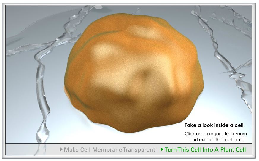
The cell in reality is very complex. The following video is a computer animation of different components in the cell. It’s based on evidence from different imaging technologies. Don’t get caught up on the technical terms! As you view the video, see if you can identify the relative sizes of different molecules or cell structures. Be as specific and detailed as possible.
ACTION
Biological Molecules in Cells
You now have a good understanding of many different uses of the 4 macronutrients. In order to describe different cellular structures it’s important to organize this information.
How could we organize the locations of these macronutrients?
Within cells, organelles perform specific tasks. This is similar to how organs perform a specific task that is important for the whole body. In organs, different tissues work together to allow it to function. On the other hand, different biological molecules work together to allow an organelle to function correctly in the cell.
Explore this interactive from Genetic Science Learning Centre about the function of organelles.
How does understanding change?
Understanding 15. What patterns do you see? Are any macromolecules more important or ubiquitous (definition:common and found in many places) than others?
Viewing Inside Cells
Biochemists have many tools available to help them to see different objects in and around cells. Some tools work well with living, or in vivo, samples. Others require special techniques only available in a lab. These samples are termed in vitro. Each tool has its advantages for the size of the structures being studied.
Imaging Technologies
Explore the following imaging technologies by clicking on each graphic in the interactive.
Select an appropriate graphic organizer to collect information about whether each tool could be used on living samples or not.
Also, connect the information to your previous work about the sizes of different cellular components: which imaging technology would be best to view them?
An effective graphic organizer will show with good details about how the size of different cellular structures are viewed by different imaging technologies.
Add this to your Portfolio. You will use this information later in this Activity.
ImagingTechnologies
How does understanding change?
Understanding 16. What can you learn from an electron microscope that you cannot learn from a light microscope?
Understanding 17. Is one type of imaging technology more useful than another when studying cell biology? Justify your response.
Experimenting on Cells
Technology is often an important consideration in an experiment. A good experiment is a fair test that should include variables that are kept the same (controlled variable) and one that is changed (independent variable). There should be a clear choice of data to be collected (dependent variable). The picture below summarizes this. You can refer to this resource page for more information about variables.
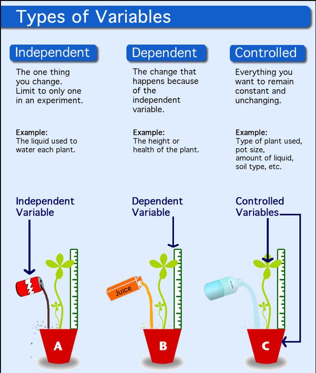
In addition to these variables, a good, fair test also includes a comparison to the experiment called a control group. (definition:a part of a fair test that is set up similar to the experiment but with known or expected results.) In the picture above, Pot C is watered with water. It is the control group because it’s used to compare the effects of the other two liquids. We can also describe it as a positive control because it’s expected that water should positively affect the dependent variable. The negative control is not shown in the picture. By contrast, a negative control would give results for no growth.
How does understanding change?
Understanding 18. Can you think of a negative control for this experiment?
 Creating a Research Plan
Consider the following fictional case:
There is a report of a new disease affecting approximately 30% of the persons living in a small rural area of Ontario. Affected individuals have a lack of energy and demonstrate a progressive loss of muscle function. Although we have no information yet, we believe the disease is caused by an infectious agent. Consequently, to limit the spread of this disease, immediate intervention is critical.
Blood and muscle tissue samples from unaffected and affected individuals are waiting for you. Microscopy and X-ray crystallography facilities are being readied for your arrival. In order to gain information as quickly as possible, please develop a solid research plan before beginning your investigations.
Some possible questions about this new disease could be:
- Is there evidence of disease at the cellular level?
- Is the disease caused by an infectious agent?
- What is the infectious agent?
- Does the infectious agent attack muscle tissue?
- How might the infectious agent cause the disease?
- Is there a drug to treat or prevent the disease?
CONSOLIDATION
Summary
- biological molecules are organized in cell structures based on their functions;
- cellular structures have properties related to the biological molecules they’re made of;
- cellular structures can be visualized in different ways depending on their size.
Investigating DNA Extraction
You are now ready to perform a lab using household materials to extract DNA from other cellular components.
Look for ways that you can present data in a clear and organized fashion, as well as practice using vocabulary related to biochemistry to describe biological molecules.
Investigation: Extracting DNA
Materials Needed:
- a sample of fruit or vegetable (such as peas, spinach, strawberries or broccoli)
- liquid dish soap
- salt
- meat tenderizer (or pineapple juice or contact lens cleaner)
- a coffee filter
- a blender or small food processor
- teaspoon
- tablespoon
- tap water
- measuring cups
- a small glass
- rubbing alcohol (70 % or more), stored in the freezer for at least 1 hour
Procedure
- Put in a blender ⅙ cup of fruit or vegetable (35 mL), a pinch of salt (less than ⅛ teaspoon), and ⅓ cup cold water (70 mL). Blend on high for 15 seconds.
- Pour your blended fruit or vegetable through the coffee filter into a small glass.
- Add ¾ tablespoons liquid dish soap (about 10 mL) and swirl to mix.
- Let the mixture sit for 5-10 minutes.
- Add a pinch of meat tenderizer to each glass and stir gently. Be careful! If you stir too hard, you’ll break up the DNA, making it harder to see.
- Tilt your glass and slowly pour rubbing alcohol into the glass down the side so that it forms a layer on top of the fruit/vegetable mixture. Pour until you have about the same amount of alcohol in the tube as fruit/vegetable mixture.
- Look for clumps of white stringy material where the water and alcohol layers meet. Recall the DNA is a long stringy molecule.
If you prefer, you can download a copy of this investigation.
Click on the button to view the rubric for this assignment.