Biological Processes and Interactions
Homeostasis: Chemical Regulation in the Body
MINDS ON
Some Key Terms
Look for these terms in this Activity. You should practice using them as you compose your responses to the upcoming tasks.
Realize, though, that this list is not exhaustive so you should look for opportunities to include other relevant terminology related to homeostasis.
| homeostasis | gland | hormone |
| pituitary gland | hypothalamus | oogenesis |
| spermatogenesis | nephron | secretion |
| pressure filtration | reabsorption | steroid |
| antagonist | osmoregulation | blood pressure |
| buffer |
Chemicals that Regulate
Look back to your graphic organizer from Unit 1, Activity 3 on Proteins, to see how chemical signals can be either hydrophilic protein hormones or hydrophobic steroid hormones. They are produced by glands located throughout the body. Unlike neurotransmitters that move signals across a synapse in the nervous system, we saw that hormones move through the blood to specific target cells. These target cells have a receptor that matches the shape of the correct hormone. Hydrophilic protein hormones have receptors on their cell membranes that send a second messenger in the cytoplasm to cause a response. Steroid hormones can pass right through the cell membrane to react with receptors inside the cell. Often these receptors, when bound to their hormone, act as transcription factors to turn on gene expression.
We have also seen how the movement of one molecule across a membrane causes the movement of another molecule. For example, in secondary active transport in the nephron, a sodium ion gradient is used to reabsorb glucose back into the blood. Other substances transported by symporter or antiporters with sodium ions include calcium ions, chloride ions, water, bicarbonate ions and protons. The protons are acidic and the bicarbonate ions are alkaline. These two ions are used in regulating the pH of the blood.
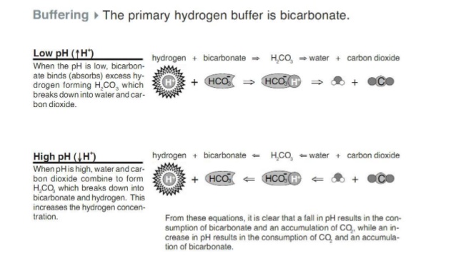
Because bicarbonate is amphoteric it acts like a buffer. In your fishbone diagram from Unit 2, Activity 2 on Enzymes, you can see that buffers help to resist changes in pH. Even though bicarbonate isn’t a hormone it still plays an important role in blood pH homeostasis.
 How do small steps lead to changes?
Change 22. How would the amount of bicarbonate in urine and respiratory rate change in response to changes in blood pH?
Glands
The concept of homeostasis has been recurring throughout this Unit. Homeostasis is important at the cellular level and is controlled by the semi-permeable cell membrane. On a population level, homeostasis is controlled by the interactions of individuals with biotic and abiotic factors. On an organism level homeostasis is controlled by several important systems. We have already examined the importance of the nervous system. Three other systems come into play here as well: the reproductive system, the excretory system and the endocrine system.
For most people, the glands (definition:an organ of the body that produces a substance that the body needs.)of the endocrine system are difficult to identify and locate in the body. Without using any other sources of information see how many glands you can correctly identify.
Glands
It’s important to know more than just where glands are located and which hormones they produce. The following video provides a good overview of the importance of the endocrine system and how it fits into homeostasis.
ACTION
Growth of the Body
Unlike the nervous system, the endocrine system responds more slowly to a stimulus. In fact, between hydrophobic and hydrophilic hormones, it’s the hydrophobic ones that take the longest time to respond because they diffuse slowly across membranes. The slowness of endocrine signalling is therefore a good fit for changes in the body leading to growth. Growth in humans occurs in three stages: infancy, childhood, and puberty.
Many hormones are important in how we grow. They are produced by two endocrine glands, the pituitary gland and thyroid, as well as the hypothalamus in the brain. In the previous video we saw that the hypothalamus acts to coordinate responses between the endocrine and nervous systems. Here, the hypothalamus coordinates metabolic activity. The stimulus could be a decrease in body temperature or it could be a drop in thyroid hormone levels in the blood. Another stimulus could be increased nutrition as we grow bigger and develop in the lead up to puberty. In all cases, hormones are secreted. (definition:the release of a substance from the interior to the exterior of a cell or organ.)
Hormones associated with the thyroid gland help regulate metabolism. One of these hormones is growth hormone, GH, (definition:a hormone secreted, in response growth hormone releasing factor, by the anterior pituitary gland and stimulates various tissues in the body.)which stimulates cells in our body to undergo mitosis. There are other thyroid hormones associated with growth and metabolism. Choose one of the following visuals to explore this further. Pay attention to different feedback mechanisms.

How do small steps lead to changes?
Changes 23. Describe how the action of thyroid hormones (T3/T4, GH) is an example of negative feedback even though metabolic rate increases.
Puberty is the last of three stages in the growth of humans, after infancy and childhood. The timing of puberty onset is still an area of scientific investigation. We do know that control over sexual maturity and production of gametes is controlled by the hypothalamus.
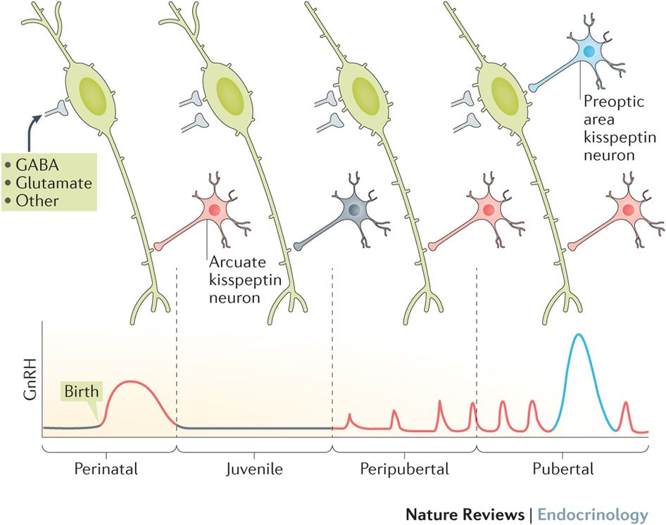
by Nature Reviews
It is generally accepted that, in general, female puberty occurs earlier than in males. Some research suggests that increased metabolic activity leads to a stimulation of the GnRH-secreting neurons. Other research indicate that with hormones produced by the gonads, (definition:the anatomical part of the body that produces gametes.)stress and seasonal changes all play a role. Transcription of dozens of genes in the hypothalamus changes at the onset of puberty. New genes are turned on which allow the hypothalamus to behave differently than during childhood. Now adolescents, like adults, are able to produce mature sperm, spermatogenesis, and egg cells, oogenesis, for the purpose of reproduction.
Just like for metabolism regulation, control over the reproductive system involves the hypothalamus and pituitary glands. Both processes use negative feedback. The target cells for pituitary hormones have different receptors depending on their biological role. For metabolism homeostasis, cells in the thyroid have cell surface receptors to TSH.(definition:a hormone secreted, in response to increased metabolic demand, by the anterior pituitary gland and stimulates the thyroid (to secrete T3 and T4 hormones). For the reproductive systems, Sertoli cells in the testes and follicle cells in the ovaries have cytoplasmic receptors to FSH, (definition:a hormone secreted, in response to gonadotropin-releasing hormone, by the anterior pituitary gland and stimulates the gonads (to secrete estrogen in females or to produce sperm in males).)follicle stimulating hormone. Production of mature gametes involves other hormones, such as luteinizing hormone, LH, (definition:a hormone secreted, in response to gonadotropin-releasing hormone, by the anterior pituitary gland and stimulates the thyroid (to secrete testosterone in males or to produce a mature egg in females).)as well as estrogen (definition:a hormone secreted, in response to FSH, by the follicle and corpus luteum and stimulates secondary female characteristics).)(sometimes also called estradiol or oestrogen) and testosterone (definition:a hormone secreted, in response to increased LH, by the Leydig cells in the testes and stimulates secondary male characteristics).)depending on sex.
We can compare feedback in the male and female reproductive systems. Even though the anatomy of reproductive systems is different, on a hormonal level there are as many similarities as there are differences. Choose one video for each sex in order to identify these details.
Male Reproductive System
Female Reproductive System
Comparing Feedback in Reproductive Systems
Summarize the differences and similarities between feedback in the male and female reproductive systems. You will use this information for an upcoming task. Your comparison should include the following details:
- structures and important cell types,
- specific hormones (FSH, LH, testosterone, estrogen, progesterone),
- examples of positive feedback,
- examples of negative feedback,
- regulation that affects the rate of gamete production.
Think about how you want to present your comparison. Ask yourself, what type of graphic organizer is best?
The following checklist will help you look at the details of your work. Your graphic organizer should:
- include an appropriate title and labels,
- present the information in a way that is easy to follow,
- present relationships that are correct and clear,
- represent an understanding of the topic and relationships among related concepts.
Blood Homeostasis
In Grade 11 we saw how the circulatory system carries nutrients and wastes around the body. Organs like the lungs, small intestines and kidneys are the sites where capillaries exchange and transport useful substances or waste products. The balance of physiological levels of these substances is also controlled through several endocrine glands.
 Canadian Contributions
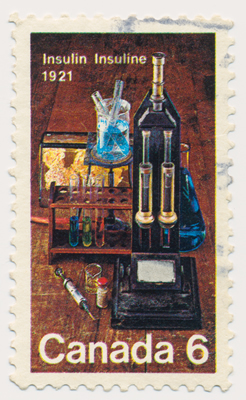
Since the 1800s doctors have known about a connection between high sugar consumption, increased urine production and the disease we now call diabetes mellitus. It was discovered that dogs with their pancreas removed would have difficulty with digestion. They also produced urine rich in glucose which attracted flies.
In the early 1900s, Canadian physicians Frederick Banting and Charles Best used extracts from the pancreas of healthy dogs to help diabetic dogs to lower their blood glucose levels to normal. The procedure was then tried using bovine insulin, the purified chemical from the pancreatic extracts, this time on humans. In 1922, the first patient was treated for this deadly disease.
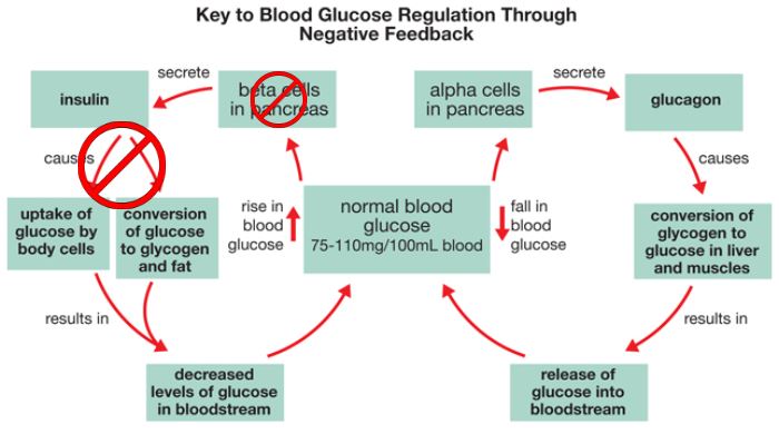
Blood glucose homeostasis is controlled by two antagonistic (definition:a biological structure or chemical substance that interferes with the action of another structure or chemical substance.)hormones, both produced by different specialized cells in the pancreas. Unlike the metabolic and reproductive hormones described earlier, glucose levels are detected by receptors in the pancreas itself. Glucagon (definition:a hormone secreted, in response to low blood glucose levels, by the pancreas and stimulates various tissues to increase blood glucose levels.)is secreted by specific pancreatic cells, called α-cells, in response to low blood glucose levels. Insulin (definition:a hormone secreted, in response to increased blood glucose levels, by the pancreas and stimulates various tissues to decrease blood glucose levels.)is secreted by specific pancreatic cells, called β-cells, in response to high glucose levels in the blood.

Glucose levels are normally regulated in response to nutrition and metabolic activities, including cellular processes that use ATP, like active transport.
How do small steps lead to changes?
Changes 24. Blood glucose regulation involves many terms beginning with the letter G. Look up the meaning of the words in the diagram to help you to keep them straight. For example, glycolysis means the breaking down of glucose: glyco- means sweet or glucose and -lysis means breaking down.
Because the same stimulus, blood glucose levels, is used for both hormones we can organize the feedback diagram in the shape of a figure eight. The stimulus is now placed in the middle of the diagram and the two hormones are represented on separate loops to the left and right from the middle.

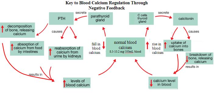
Another physiological condition controlled by antagonistic hormones is blood calcium homeostasis. In this case the two hormones are secreted by two different glands: the thyroid and the parathyroid.
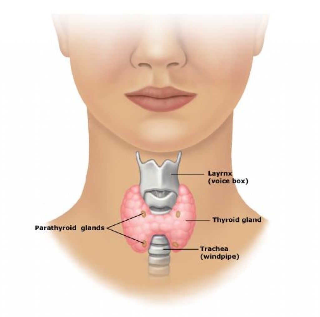
Again the stimulus is detected by receptors in the gland rather than by the peripheral nervous system. Parathyroid hormone, PTH, (definition:a hormone secreted, in response to low blood calcium levels, by the parathyroid glands and stimulates various tissues to increase blood calcium levels.)is secreted by the parathyroid in response low blood calcium levels. Calcitonin (definition:a hormone secreted, in response to increased blood calcium levels, by the thyroid and stimulates various tissues to decrease blood calcium levels.)is secreted by the thyroid in response to high calcium levels in the blood. Calcium levels are normally regulated in response to nutrition and cellular processes, including the release of neurotransmitters by neurons and muscle contractions.
Similar to the feedback diagram involving insulin and glucagon, blood calcium levels can be organized in a figure eight shaped feedback diagram. The effects of each hormone target different cell types leading to us to show branches off each feedback loop.
How do small steps lead to changes?
Changes 25. In humans, both calcium and glucose are obtained from our diet and are absorbed by the small intestine. One of the issues related to food systems in developed countries like Canada is overnutrition of essential nutrients. Compare how blood glucose and blood calcium levels are regulated when there is a surplus of each nutrient in the body. Identify both similarities and differences in how calcium and glucose are stored and excreted by the body.
Doctors studying diabetes mellitus patients noticed that their urine contained glucose and that their blood glucose levels were higher than normal. For these patients, excess glucose was being removed by the kidney in urine. Similarly, high levels of blood calcium are removed by the kidney, although this process occurs in healthy people.
Refer back to your flowchart from Unit 2, Activity 3 on Cellular Transport, to see how the structure in the kidney that is responsible for cleaning the blood is the nephron. We also saw that substances move both into and out of the nephron. All together there are four steps to urine formation by the nephron: filtration, reabsorption, secretion, and excretion. Watch the following video to learn more about where these processes occur within the structures of the nephron.
Mapping out Urine Formation
As you continue to explore this section of this Activity you will be mapping out the steps of urine formation in a flowchart. At this point we will start working on that flowchart. Your flowchart should include these details and concepts:
- location in the nephron where the reactions occur,
- the types of substances moving into or out of the nephron.
Be sure to leave room for more details to add as you work through this Activity.
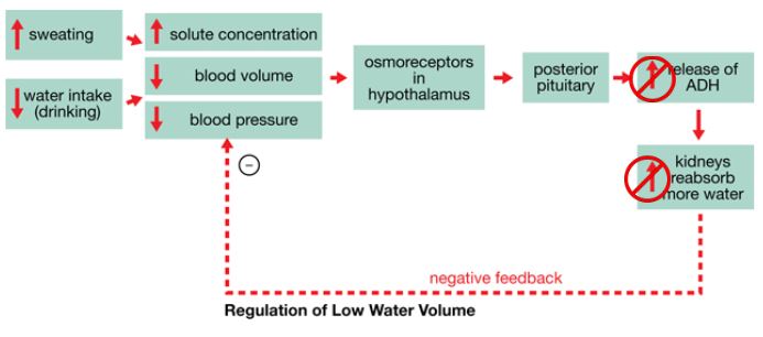
In the case of glucose, in healthy people glucose is both filtered and then reabsorbed at the proximal convoluted tubule. Diabetics do the same, but they reabsorb a smaller percentage of glucose. Similarly, calcium is filtered and reabsorbed, although here, the reabsorption happens at the distal convoluted tubule. PTH helps increase the permeability of the distal convoluted tubule to allow for a greater reabsorption of calcium if blood calcium levels are low. Shortly we will learn about another hormone, antidiuretic hormone, or ADH (definition:a hormone secreted, in response to low blood pressure, by the posterior pituitary gland and stimulates the collecting duct to become more permeable to water reabsorption.)that helps to reabsorb excess water at the collective duct.
Most importantly, the kidneys are responsible for water homeostasis. Too much water excreted in urine leaves us dehydrated: unable to effectively move solutes around the body and unable to regulate our internal body temperature. Too little water excreted in the urine leaves too much water in the blood.
Why is shape important in Biology?
Shape 23. The length of the loop of Henle is different in different species. Why might its length be an adaptation that helps animals to survive in hotter or drier environments?
High blood volume causes the heart to work harder to circulate the blood around the body. The result is an increase in blood pressure. In the short term, increases in blood pressure are useful especially for short bursts of energy. In the long run, however, high blood pressure stresses out the circulatory system. The risk of blood vessels bursting increases as does the frequency of forming blood clots and these effects can cause a heart attack or stroke in the brain. Blood pressure must therefore be carefully regulated by the body.
Hormones act on the nephron to regulate blood pressure. There are actually four main hormones that act to regulate blood pressure. For our purposes in this course we will focus on two of them: aldosterone (definition:a hormone secreted, in response to high osmotic pressure, by the adrenal gland and stimulates the nephron to reabsorb more Na+ ions from the filtrate) and ADH.
Map of Urine Formation
As you watch this next video, look for the direction that water moves in the nephron. Add details to your Map of Urine Formation. Which of the four steps in urine formation involve aldosterone and which involve ADH?
In the story of blood pressure regulation we can see that water is moving. This happens through aquaporins in cell membranes by osmosis which is against a solute concentration gradient. You can appreciate that in order to properly regulate blood pressure the kidney must also be able to properly regulate the solute concentration gradient. This regulation is called osmoregulation. The main solute nephrons transport in order to establish a concentration gradient is the sodium ion using sodium potassium pumps or facilitated diffusion.
As you watch the following video, look for the three places that sodium ions are moved in the nephron in order to establish a hypertonic concentration gradient. Identify if this movement is active transport or passive transport.
How do small steps lead to changes?
Changes 26. Both ADH and aldosterone are hormones that act on the nephron and help with water movement. Find other similarities and differences as you compare these two hormones. Could terms like “permeable” and “hypertonic” help to add good details to your answer?
 Homeostasis
What did you learn about the uses of homeostasis?
3 - Three things I learned about how homeostasis is used in the body:
2 - Two other things I found interesting about homeostatic processes:
1 - One question I still have:
Your flowchart should include these concepts and details:
- location in the nephron where the reactions occur,
- the types of substances moving into or out of the nephron,
- vocabulary and terminology that allow you to demonstrate your understanding.
Ask yourself these big questions as a way to monitor and improve your work:
- Is the information I have presented a complete representation of my learning?
- Is the information I have presented correct?
- Have I fulfilled the requirements of the task?
- Are the steps I describe organized in the correct order?
Medication Side Effects
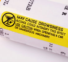
Modern medications have had a powerful impact on the health outcomes of human populations. Part of the process of developing new medicines is its testing to identify an effective dose and possible side effects. (definition:any effect of a drug or medication other than its intended effect.) Many drugs work on a variety of cell types because they lack specificity like hormones. Other drugs directly interfere with the function of chemical signals in the body. In both cases, medications can have unintended side effects. Pharmaceutical companies warn patients of these side effects through special labels or on the side of medication packaging.
 How do small steps lead to changes?
Try searching for a medication that has a side effect that interferes with the proper functioning of a hormone. This could affect either the endocrine system, reproductive system or excretory system. What is the medication supposed to be used for? Explain how normal hormone feedback changed.
Diagnosing Disorders
An imbalance in the concentration gradient in the kidneys can change the amount of water reabsorbed by the nephron. For example, diabetes patients produce large amounts of urine. In the case of diabetes mellitus, as more glucose stays in the filtrate in the urine this leaves more solutes than usual.
This in turn lowers the concentration gradient. As a result, less reabsorption by osmosis happens and more water stays in the urine.
Another type of diabetes is caused by a deficiency in ADH signalling. Here, less water is reabsorbed because the collecting duct stays impermeable to water. Again, as a result, more water stays in the urine. This type of diabetes is called diabetes insipidus and is not related to glucose homeostasis.
How do small steps lead to changes?
Let’s take a look to see how feedback diagrams can help us to explain the symptoms of a disease and help doctors to recommend treatments. Search for a disorder caused by a change in hormone (definition:TSH, TRH, thyroxine, GH (somatotropin), GnRH, FSH, LH, estrogen, testosterone, progesterone, insulin, glucagon, calcitonin, PTH, aldosterone, ADH.)signalling. Perhaps there is a disorder in which the hormone is overproduced, or maybe it’s underproduced or not secreted at all. Other diseases could be caused by an inability for the receptor to recognize the hormone. Choose a disorder related to a hormone that you can describe with the best and most specific details.
Find the following details about the disorder you are researching:
- the gland that secretes the hormone,
- at least one symptom of the disorder,
- a physiological measurement used by doctors in diagnosing the disorder.
 Self-Check Quiz
CONSOLIDATION
Summary
- the endocrine system coordinates long-term homeostasis in the body;
- growth and metabolism in humans is controlled through negative feedback originating at the pituitary gland;
- blood homeostasis is controlled by endocrine glands and the kidneys;
- changes to endocrine homeostasis can lead to disorders that affect the health of patients.